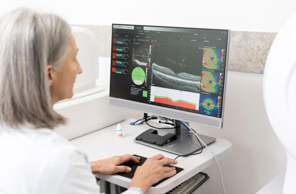
If it’s been a while since you’ve sat in the comfort of your optometrist’s exam chair, it might surprise you to know just how far the technology we use in routine eye exams has come. Take OCT scans, for example.
This imaging tool takes detailed scans of the retina’s layers, uncovering eye conditions such as glaucoma, macular degeneration, and even drug-related retinal changes, often before you notice any symptoms.
The OCT Scan, Explained
The right tools can make a noticeable difference in the quality and experience of your routine eye exam. With optical coherence tomography, or OCT for short, we can uncover fine details of your eye’s internal structures, making it an integral part of your visit.
An OCT scan is a noninvasive test that uses light waves to take detailed, cross-sectional images of your retina, the light-sensitive tissue lining the back of your eye.
Although we focus on one eye at a time, the process is simple:
- Rest your chin and forehead against the machine
- Gaze at the focal point
- Try to keep your eyes open
Within seconds, the machine captures valuable information about your visual health: no drops, no discomfort, and no fuss. A few moments are often all it takes.
What OCT Scans Can Detect
The brilliant thing about OCT scans is that they can capture fine details, detecting any abnormal changes within the retina, especially major eye concerns that pose a threat to your vision.
Macular Degeneration
Age-related macular degeneration (AMD) is one of the leading causes of vision loss for older adults. An OCT scan can detect the earliest signs of AMD, often before you notice any vision changes. The scan shows detailed images of the macula, the part of your retina responsible for sharp, central vision.
OCT technology can detect fluid buildup, abnormal blood vessel growth, and changes in the retinal pigment epithelium that indicate the development of AMD. Early detection means earlier treatment options and better outcomes for preserving your central vision.
Diabetic Retinopathy
If you have diabetes, regular OCT scans are incredibly valuable for monitoring your eye health. Diabetic retinopathy (which often develops without noticeable symptoms) occurs when high blood sugar damages the tiny blood vessels in your retina.
An OCT scan can reveal swelling, fluid leakage, and other retinal changes that may not be visible with standard eye exam technology. This scan helps your optometrist track the progression of diabetic eye disease and determine the right course of treatment.
Glaucoma
Glaucoma refers to a group of eye diseases that damage the optic nerve, a structure responsible for strong peripheral (side) vision. Although glaucoma has several risk factors, elevated eye pressure is a major risk factor and often contributes to optic nerve damage.
The challenge with glaucoma is that it typically manifests and progresses without symptoms until significant optic nerve damage occurs.
This is where OCT scans can make a difference in glaucoma detection and monitoring. They can measure the thickness of your retinal nerve fiber layer and identify changes before vision loss becomes noticeable. Not to mention, OCT scans can help optometrists distinguish between normal age-related changes and damage related to glaucoma.
Macular Holes and Pucker
Sometimes the delicate tissue of your macula can develop holes or wrinkles that affect your central vision.
OCT scans detect these conditions by providing cross-sectional views that show the exact location, size, and severity of macular holes or epiretinal membranes (macular pucker). This helps determine whether surgical intervention is necessary, and allows your optometrist to monitor changes over time.
Retinal Detachment
A retinal detachment refers to when the retina pulls away from the underlying tissue—no doubt, a serious eye concern that requires immediate intervention. OCT scans can detect subtle detachments that might be missed during routine examination.
If you notice flashes of light or an increase in floaters, visit your optometrist right away for a thorough evaluation. Delaying treatment can pose a threat to your vision.

Less Common, But Important Concerns
Though these eye conditions are less common, an OCT scan can also detect:
- Central serous retinopathy: A condition that involves fluid buildup under the retina. OCT scans can clearly show the fluid accumulation and help track whether it resolves on its own or requires treatment.
- Retinal vein occlusions: When blood vessels in your retina become blocked, it can cause vision problems and retinal damage. OCT scans can detect the swelling and fluid buildup associated with these blockages, helping guide appropriate treatment.
- Drug-related retinal changes: Some medications can affect your retina over time. OCT monitoring allows your optometrist to detect these changes early and coordinate with your primary care physician or specialist to make any necessary adjustments.
OCT Scan vs Fundus Photos
Retinal imaging, such as OCT scans and fundus photography (2 different technologies), is a valuable tool for diagnosing and monitoring eye health.
OCT scans capture detailed images of the layers of your retina, while fundus photos provide a full-color picture of your eye’s internal structures. This includes the optic nerve, blood vessels, and the retina.
Though the details of these images differ, together they can provide your optometrist with a comprehensive picture of your visual health.
- Identifying potential concerns early: OCT scans and fundus photos complement each other to catch potential problems such as glaucoma or retinal tears early.
- Monitoring over time: With routine imaging, we can use these tools to track any changes or progression over weeks, months, or years. Your optometrist can compare images side by side, spotting tiny changes that could be early signs of progression in conditions like diabetic eye disease.
- Adjusting management plans: If you’re undergoing treatment (perhaps for macular degeneration or glaucoma), these technologies can show how your eyes are responding. These tools guide your optometrist in fine-tuning your treatment plan so it fits your needs perfectly.
Take Charge of Your Eye Health
We value technical innovations because of how easily they can improve our lives. Vision care is no different. Before OCT scans, many retinal conditions could only be detected when they were already quite advanced, and in turn, more challenging to manage. With OCT scans and fundus photos, we can identify problems earlier to facilitate better outcomes.
Our comprehensive eye exams include retinal imaging for this reason! Combining technology with professional care can help maintain your eyes’ health and keep your vision sharp. Connect with our Bethany Eye Care team to book your routine eye exam!


